Tuesday, November 15, 2011

The Lasting Impression

Friday, on the final day of visiting the MicroAquarium, the changes in population I observed were minimal in comparison to previous visits. It would seem that this stage has been reached because each organism has found a bit of a niche for itself to fill. The diatoms remained mostly at the bottom while the Actinosphaerium patrolled the middle range with the most of the older rotifers. I did notice several new, more aggressive rotifers, Rotifera philodina (Raims and Russell 1996 p. 188). They were busily sensing for and consuming smaller protists and rotifers in and around the plant material. Vorticella, centered in the picture to the right, was new to me as well and a very interactive organism (Raims and Russell 1996 p. 104). When I bumped the tank accidentally, their bodies floating out on long stem-like structures, quickly retracted to the leaf material they were attached to. There were about five of them were interspersed along a branch of the moss and large groups of ten or so amassed along the bottom.


Rotifera

Also dotting the middle layer, several dark brown Difflugia camped around detritus (Raims and Russell 1996 p. 160). The uppermost layer was dominated by quick-swimming Cohnilembus (Pennak 1953 p. 66 Fig. B) and young rotifers still working on the food that had been added to the tank.

 Difflugia


 Raims KG. Russell BJ. 1996. Guide to Microlife. Canada. Franklin Watts. 104-188 p.

Thursday, November 10, 2011

A Delicate Balance

Upon visiting my MicroAquarium Friday, I observed a very dead Utricularia gibba L. still drifting in the medium. Considering that the other plant was still thriving, I assume the carnivorous action of this plant was unsuccessful in capturing enough nutrients for the plant's survival. I recall sighting only a few organisms that had been lured into its trap on previous visits. Otherwise to the naked eye, my tank looked very much the same. I needed to add a little more water again, and I did note that a second food pellet had been added. The diatoms that I had been following at first appeared to be gone as well, but near the end of my observation time I located hundreds of them gathered at the bottom of the MicroAquarium, near the sediment. They were looking much more diverse than ever before, donning at least twenty new morphologies. Unfortunately, I was unable to view most of them close enough to make out significant detail even under high magnification due to their incredibly smaller size. Now, I am only hoping to see more growth out of them.


 Diatoms

 Aside from the beautiful silica-shelled diatoms, the other organisms had been flourishing as well. Many I have become familiar with in the tank such as the crustacean-like rotifers, Euplotes, Actinosphaerium with all its pins and Stylonychia mytilus bearing its narrow posterior accented by just a few waving cilia were still active in the scene and in assorted sizes, unlike the various tiny diatoms. The Actinosphaerium numbers had at least doubled. Rotifer eggs, it seems were not in as plentiful supply as my last visit. The same could also be said of the rotifers themselves. Though the difference was minimal this time, we'll see how their lower numbers play out in the future.

A few new faces were scurrying beneath my lens on Friday as well. A very quick-moving Cohnilembus (Pennak 1953 p. 66 Fig. B) and his cronies were spotted several times, although all photograph attempts were unsuccessful. They were easily recognizable because of the numerous cilia that followed the outside rim of their body, capped by a single caudal cilia. I found their movements knife-like at times. Another newly identified individual was a charming Tachysoma (Patterson 1996 p. 125 Fig. 264-265) who fooled me for a Paramecium at first under the 4x lens. The grouping of cilia at the anterior and what appeared to be the posterior ends poised it distinctly different to my untrained eye. Once I had identified one, I noticed several flitting about between the muck and the detritus. Their numbers were hard to estimate considering their speed and adroit evasiveness.


Pennak RW. 1953. Fresh-water Invertebrates of the United States. New York. The Ronald Press Company. 64-67 p.
Patterson DJ. 1996 Freeliving Freshwater Protozoa. London. Manson Publishing LTD. 264-265 p.

Tuesday, November 1, 2011

The Race for World Dominance

Only two weeks into the experiment, and the growth is astounding. I imagine this is owed mostly to the decomposing food matter I identified that had been introduced to the MicroAquarium. I extracted the details from the class blog: "Atison's Betta Food" added October 21 made by Ocean Nutrition, Aqua Pet Americas, 3528 West 500 South, Salt Lake City, UT 84104. Ingredients: Fish meal, wheat flower, soy meal, krill meal, minerals, vitamins and preservatives. Analysis: Crude Protein 36%; Crude fat 4.5%; Crude Fiber 3.5%; Moisture 8% and Ash 15%. Where previously locating an organism to view required a few seconds of probing, now tens of organisms were competing for food and space in any given section. Almost all of the organisms were many times smaller than a millimeter and much more diverse. Some of the same organisms were present. A few I had not identified before such as Uroglenopsis (Pennak 1953 p. 41 Fig. G), Enteroplea lacustris (Pennak 1953 p. 200 Fig. E), Paramecium (Pennak 1953 p. 25 Fig. E) were still thriving. I was able to even identify the large green sacs that I had noticed unmoving and scattered around the aquarium last time as rotifer eggs (Pennak 1953 p. 174 Fig. A). In addition, the number of diatoms had significantly multiplied, but the varieties were relatively the same. A common one looked very similar to the rice-shaped one from last week, but with a budding protuberance on each narrow end.


 Actinosphaerium


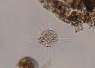
The first new organism I discovered feeding near the now deceased carnivorous plant was Euplotes (Pennak 1953 p. 69 Fig. B). While I was observing him he swam right into the most interesting looking spherical organism with long flagella, Actinosphaerium (Pennak 1953 p. 57 Fig. C). A plethora of organisms were gathered in the area encompassing the added food. One such organism was Holosticha algivora (Bick 1972 p. 19 Fig. 8C), drifting on the outskirts. Others I observed gathered near the feeding frenzy were  Stylonychia mytilus  (Bick 1972 p 174-175 Fig 88) and Thuricola (Pennak 1953 p. 72 Fig. H). There were also many more rotifer eggs dispersed throughout this trafficked section at the top of the MicroAquarium.


Pennak RW. 1953. Fresh-water Invertebrates of the United States. New York. The Ronald Press Company. 25-200 p.
Bick H. 1972. Cilated Protozoa. Geneva, Switzerland. World Health Organization. 19-175 p.

Monday, October 24, 2011

A Glimpse of Change

On Friday, I visited my MicroAquarium for the first time. The seemingly isolated life in the sample of quarry water I had started with had proliferated magnificently in just little over a week's time. I found myself fascinated by the observation of these mostly microscopic creatures. At first, the aquarium had appeared just the way I had left it, but when I turned it over to place under the microscope lens I noticed the furious activity of a water flea whom had been displaced. He darted into the mud at the bottom and I did not see him again. Upon closer inspection with the aid of the microscope, I was able to identify the feeding activity of many Paramecium  mostly on the dead leaf material that I had added at the end of my last session with the aquarium, but I am still working on uncovering the specific species. I was a little disappointed to note that even with scouring I was only able to uncover a single rice-shaped brown diatom about 11-12mm in length. There were also several organisms 7-10mm in length that were crawling through the layers of the leaf material but I could not glimpse them sufficiently for identification either.

Saturday, October 15, 2011

Creating the MicroAquarium

  This week we all selected a source of water from which to draw upon for our MicroAquariums. I selected the water and sediment sample from Mead's Quarry, one of the larger bodies to choose. The color and murkiness was rather average, but I'm hoping to find a lot of interesting kinds of little life in it. I am familiar with the area myself, so it will be exciting to see up close the kinds of creatures I have walked past.


  In order to get an accurate re-creation, the tiniest bit of sediment was added across the bottom of the aquarium to start. This ensures that I have a media for the plants I will be adding to grow and flourish in. Then I extracted some water from the bottom of the water source container and carefully dripped it until the MicroAquarium was a third full. The second third of water was taken from the middle of the container, and the final from across the very top. Such meticulous water sampling allows for the myriad organisms inhabiting the various layers to hopefully join me in my aquarium for observation.
Then, we each added a snippet of Amblestegium sp. moss collected from a natural spring at Carter's Mill Park and Utricularia gibba L., a carnivorous, flowering plant grown in water tanks outside of greenhouse at Hesler Biology Building to the mix. Plants are important as they will create a natural water filter and supply the various organisms with the nutrients and requirements they need to survive and thrive.
   After a few adjustments, I was ready to view my MicroAquarium under the microscope. However, my neighbor was much further along and happened to capture a little diatom in his! I've previously seen pictures of them, and have always been very interested. Dr. McFarland noticed my enthusiasm and suggested that I add a small piece of leaf into my aquarium as well to promote the growth of more diatoms in my own. I am eager to see how this maneuver turns out in the upcoming weeks.

  In my own aquarium I observed much life as expected, but I could not identify any organisms at the time. The bulk of the life I saw was floating in the open middle of my setup, and all seemed single-celled and unmoving. A few had a green or blue hue to them, but most were colorless.